Recognizing Potentially Malignant Oral Disorders and Oral Cancer
By Tiffany Tavares, D.D.S., and Alessandro Villa, D.D.S., Ph.D., MPH
 Oral cancer (OC) is an important public health concern, with an estimated 32,580 new cases in the United States in 2017 and 7,550 deaths with an average five-year survival rate of 64.5% (1). However, this rate drops to 38.5% in cases of distant disease, in contrast to 83.7% when the disease is localized (1). In addition to better survival rates, patients with early-stage disease generally have fewer sequelae from treatment (surgery alone vs. radiation and/or chemotherapy) (2, 4).
Oral cancer and oral potentially malignant disorders: the role of the dentist
Meticulous inspection of all the soft and hard tissues of the oral cavity should be routine practice — not only to detect oral potentially malignant disorders (OPMDs) or OC, but also any other disease in the oral cavity (e.g. vesciculous bullous disorders, benign lesions, immune-mediated conditions, etc.).
A single-center study of 51 patients with a new diagnosis of oral and oropharyngeal squamous cell carcinoma (SCC) showed that nearly one-third of these patients had been referred by a dental provider; they were less likely to have presented with symptoms and had a lower stage than those referred by medical physicians (4). From a medico-legal standpoint, a 2015 study that reviewed three United States legal databases found 41 cases in which lawsuits were filed against dental practitioners for alleged head and neck, oral, or oropharyngeal cancer misdiagnosis or diagnostic delay with the highest recovery amount of $3.5 million and a mean of $1.03 million for the plaintiffs (5).
What can we do?
Medical and Family History
A thorough review of the patient’s medical and family history is important to identify individuals with risk factors for OC, such as:
Modifiable risk factors:
- Current and former tobacco use (especially smoking): opportunity for cessation counseling where applicable
- Areca nut chewing
- Heavy alcohol consumption
- UV light (for lip cancer)
Medical history:
- Personal and/or family history of cancer (especially head and neck cancer)
- History of radiation therapy to the head and neck
- Persistent HPV infection (for oropharyngeal cancer)
- Autoimmune diseases and/or prolonged immunosuppression
- Graft-versus-host disease
- Plummer-Vinson Syndrome
Geriatric predisposition:
- Fanconi anemia
- Dyskeratosis congenita
- Epidermolysis bullosa (6-8)
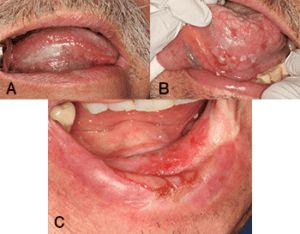
Figure 1 |
|
| A – Homogenous leukoplakia on the left ventrolateral tongue. |
| B – Non-homogenous leukoplakia with areas of ulceration on the right ventrolateral tongue. Note the fissuring on the more hyperkeratotic patches. |
| C – Erythroplakia with dysplasia on the anterior mandibular alveolar ridge in a patient with a history of oral squamous cell carcinoma after a composite resection and radiation therapy. |
Physical Examination
- A thorough extraoral examination is important to identify:
- Cervical lymphadenopathy (typically firm and fixed)
Trismus and facial asymmetry; this may indicate infiltration of muscle and other adjacent structures (6).
Visual and tactile intraoral examination is the most important component for the detection of OPMD and OC as pain and other symptoms such as numbness, hoarseness or dysphagia, are infrequently present unless the patient has advanced disease (3, 6). High-risk sites are the ventral tongue and floor of mouth (6-8).
Clinical Presentation
OMPD such as localized leukoplakia (LL), erythroplakia and proliferative leukoplakia (PL) may present as white, red, or mixed red/white plaque. Leukoplakia may be homogenous, with or without fissuring, or non-homogenous with nodular or verrucous features and associated erythema. Non-homogenous lesions have a higher risk of transformation (7, 8).
OC may present with all the same features as OMPD in addition to the following:
- Speckled changes
- Nodules or masses
- Ulceration (the presence of rolled borders is concerning for OC)
- Induration (7, 8)
OMPD and OC may be misdiagnosed on clinical examination as reactive or inflammatory lesions such as frictional keratosis (e.g., bite trauma, benign alveolar ridge keratosis), traumatic ulcerations, contact desquamation, and lichen planus, among others (7). As such, any lesion of unclear etiology that has not responded to treatment/intervention nor resolved in two weeks should elicit suspicion and be promptly biopsied. If the dental practitioner is not comfortable in performing a biopsy or is unsure of the best area to sample, a referral to an oral medicine specialist or oral pathologist or OMFS may be helpful (7, 8). In cases of non-homogenous lesions, more than one biopsy may be necessary to assess the different areas involved (7, 8).
Not all leukoplakias are the same
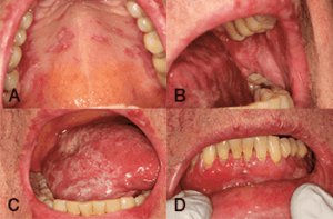
Figure 2 |
|
| Proliferative erythroleukoplakia characterized by multifocal mixed red and white patches and plaquesinvolving the hard palate, buccal mucosa, ventrolateral tongue and gingiva in the same patient. Of note, these lesions were on the bilateral buccal mucosa and bilateral ventrolateral tongue. This does not represent oral lichen planus. |
True leukoplakia is essentially a diagnosis of exclusion based on clinical appearance and medical history with histopathologic confirmation (7). LL has an overall malignant transformation rate of 3 to 15% and an annual transformation rate of one to three% (7-9). Upon biopsy, 20 to 40% of leukoplakias represent dysplasia, carcinoma in situ or SCC, in contrast to 90% in erythroplakias (7, 8). LL and erythroplakia are more commonly seen in men and associated with smoking (7, 8). Another distinct entity to be aware of is PL, an aggressive form of leukoplakia with multifocal presentation, more commonly seen in females; PL is not strongly associated with smoking, and has a 70 to 100% malignant transformation rate (over 10 to 20 years), although less than 10% of these demonstrate dysplasia or SCC at first biopsy (7-9).
Important features to note on biopsy reports are the presence carcinoma, and also the presence and severity of dysplasia and whether this extends to the tissue edges (7-9).
Treatment for OMPD and follow-up for OMPD and OC
Treatment options for OMPD include wide excision, laser ablation, or watchful waiting (9, 10). Follow-up schedules vary based on the presence and severity of dysplasia, history of OC and time from OC treatment and typically range from three to 12 months. Oral medicine specialists, oral pathologists, ENTs and OMFS may be involved in following these patients (7-9).
For extensive lesions, excision is not always an option, and, unfortunately, there are no good evidence-based preventive agents or nonsurgical approaches (although available) to prevent malignant transformation (10). There are also no biomarkers to indicate which lesions are more likely to progress to aid treatment decisions (9, 11). The currently available marketed diagnostic aids/screening tools for detection of OC and OMPD are not recommended for general practitioner use due to low specificity, high-risk of bias, and lack of well-designed prospective, randomized control trials supporting their use (11).
As such, close follow up with consistent photographic documentation is imperative, at initial examination and every follow-up to monitor for alterations, with a low threshold for re-biopsy. The patient should be advised to return promptly should there be any changes in texture (nodularity, ulceration, increased keratosis and/or erythema) or size, or development symptoms (such as pain, numbness, etc.).
Dr. Tiffany Tavares is the chief resident and a DMSc candidate in the Oral Medicine Residency program at Brigham and Women’s Hospital/Harvard School of Dental Medicine. Dr. Tavares obtained her D.D.S. from Brazil, where she developed an interest for oral premalignancy and oral cancer. Dr. Tavares received a grant from the American Academy of Oral Medicine for her doctoral research project on combination drug therapy in head and neck squamous cell carcinoma. Dr. Tavares can be reached at ttavares1@partners.org.
Dr. Alessandro Villa is an associate surgeon in the Division of Oral Medicine and Dentistry at Brigham and Women’s Hospital/Harvard School of Dental Medicine, where he also serves as the Program Director of the Oral Medicine Residency program. Dr. Villa obtained his D.D.S. degree and a Ph.D. from Italy. He served as a post-doctoral fellow at the National Cancer Institute, where he studied the oral HPV prevalence in a large population and methods of early detection of oral cancer. He obtained his Master of Public Health from AT Still University, Missouri, and his Certificate in Oral Medicine from the BWH/HSDM. Dr. Villa’s research interests are focused primarily on leukoplakia and oral complications from cancer treatment.
References |
| 1. Siegel, R. L., Miller, K. D. and Jemal, A. (2017), Cancer statistics, 2017 CA Cancer J Clin. 2017;67:7–30. |
| 2. Stefanuto, Peter, Jean-Charles Doucet, and Chad Robertson. Delays in treatment of oral cancer: a review of the current literature. Oral Surg Oral Med Oral Pathol Oral Radiol. 2014;117:424-9. |
| 3. Dolan, Robert W., Charles W. Vaughan, and Fuleihan Nabil. Symptoms in early head and neck cancer: an inadequate indicator. Otolaryngol Head Neck Surg. 1998;119:463-7. |
| 4. Holmes, Jon D., et al. Is detection of oral and oropharyngeal squamous cancer by a dental health care provider associated with a lower stage at diagnosis J Oral Maxillofac Surg. 2003;61:285-91. |
| 5. Epstein, Joel B., et al. Head and neck, oral, and oropharyngeal cancer: a review of medicolegal cases. Oral Surg Oral Med Oral Pathol Oral Radiol. 2015; 119:177-86. |
| 6. Chi, Angela C., Terry A. Day, and Brad W. Neville. Oral cavity and oropharyngeal squamous cell carcinoma — an update. CA Cancer J Clin. 2015;65:401-21. |
| 7. Warnakulasuriya, S., Johnson, N.W., and Van der Waal, I. Nomenclature and classification of potentially malignant disorders of the oral mucosa. J Oral Pathol Med. 2007;36:575-80. |
| 8. Villa, A. and Woo, S-B. Leukoplakia — A Diagnostic and Management Algorithm. J Oral Maxillofac Surg. 2017;75:723-34. |
| 9. Woo, S.B., Grammer, R.L., Lerman, M.A. Keratosis of unknown significance and leukoplakia: A preliminary study. Oral Surg Oral Med Oral Pathol Oral Radiol. 2014;118:713. |
| 10. Lodi G, Franchini R, Warnakulasuriya S, Varoni EM, Sardella A, Kerr AR, Carrassi A, MacDonald LCI, Worthington HV. Interventions for treating oral leukoplakia to prevent oral cancer. Cochrane Database Syst Rev. 2016;7:CD001829 |
| 11. Macey R, Walsh T, Brocklehurst P, et al. Diagnostic tests for oral cancer and potentially malignant disorders in patients presenting with clinically evident lesions. Cochrane Database Syst Rev. 2015;5:CD010276. |


